
When Every Minute Counts, the Right Imaging Technology Makes All the Difference
Something is wrong inside your body. You can feel it. But what exactly? And how serious is it? This is the moment when the diagnostic tools available to your emergency care team become absolutely critical. In emergency medicine, the ability to see inside the body quickly and accurately, without surgery, can be the difference between life and death.
CT scanning is one of the most powerful diagnostic tools in modern emergency medicine. It provides detailed, three-dimensional images of every internal structure, bones, organs, blood vessels, and soft tissues, within minutes. Understanding what a CT scan is, how it works, when it is needed, and where to access one in Houston gives you the knowledge to make fast, informed decisions when a medical emergency strikes.
What Is a CT Scan?
A CT scan, short for computed tomography, is an advanced medical imaging technology that uses a combination of X-rays and sophisticated computer processing to produce highly detailed, three-dimensional images of structures inside the body.
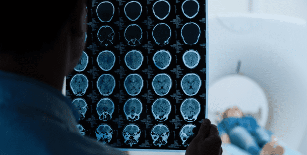
Unlike a standard X-ray that captures a single flat image from one angle, a CT scanner rotates around the patient and captures hundreds of images from multiple angles simultaneously. A powerful computer then assembles these images into detailed cross-sectional “slices”, and stacks those slices into a comprehensive three-dimensional reconstruction of the scanned area.
The result is a complete, nuanced visual map of the body’s interior, showing bones, muscles, organs, blood vessels, and abnormal structures like tumors, clots, or fluid collections, all without a single incision.
CT scanning is considered one of the most transformative technologies in the history of emergency medicine. It allows physicians to diagnose conditions in minutes that would previously have required exploratory surgery, dramatically improving both speed of diagnosis and patient outcomes.
How Does a CT Scanner Work?
Understanding the mechanics of CT scanning helps demystify the experience for patients who may feel anxious about the procedure.
The Physical Process
A CT scanner consists of a large, ring-shaped machine called a gantry, the distinctive donut-shaped structure you lie inside. Inside this ring, an X-ray source and a digital X-ray detector face each other on opposite sides of the ring.
Here is what happens during a scan:
Step 1: The patient lies on a motorized examination table that slides slowly through the center of the gantry ring.
Step 2: The X-ray source rotates continuously around the inside of the gantry, completing full 360-degree rotations as the table moves through the machine.
Step 3: As the X-ray source rotates, it fires narrow beams of radiation through the body from hundreds of different angles.
Step 4: The digital detector on the opposite side of the ring captures the X-rays that pass through the body. Different tissues absorb different amounts of radiation, creating varying signal intensities at the detector.
Step 5: The computer receives the data from the detector and processes it in real time, reconstructing cross-sectional images called slices. Each slice represents a thin cross-section of the body at that position.
Step 6: The slices are stacked by the computer into a three-dimensional volume, allowing radiologists and emergency physicians to examine any plane, any angle, and any structure within the scanned area.
Step 7: The final images are available for physician review within minutes of the scan completion, providing fast, actionable diagnostic information.
What the CT Scan Can Detect That Standard X-Rays Cannot
The CT scanner’s rotating, multi-angle approach gives it access to hundreds of different tissue density levels, far beyond what a standard X-ray can differentiate. While an X-ray distinguishes primarily between dense structures (bone, metal) and less dense structures (soft tissue, air), a CT scanner can detect subtle density differences within solid organs, identifying inflammation, early tumors, fluid collections, and tissue damage that would be completely invisible on standard X-ray.
CT Scan vs. Standard X-Ray
Both technologies use X-ray radiation and are valuable in emergency medicine. But they serve fundamentally different diagnostic purposes. Choosing the right one, or using both appropriately, depends on what the physician needs to see.
Standard X-Ray
A standard X-ray uses a single, fixed radiation beam that passes through the body from one direction and creates a flat, two-dimensional image on a detector plate.
Best for:
- Identifying bone fractures and dislocations, calcium in bones blocks radiation effectively, creating sharp contrast
- Detecting pneumonia or fluid in the lungs, visible as haziness on a chest X-ray
- Identifying foreign objects like swallowed items or embedded fragments
- Initial screening for major structural problems
- Quick, low-cost imaging when detailed soft tissue evaluation is not required
Limitations:
- Cannot show subtle bone injuries or stress fractures
- Poor soft tissue differentiation, internal organs appear as undifferentiated gray shadows
- Single-plane imaging only, cannot show depth or provide three-dimensional context
- Cannot reliably detect tumors, abscesses, vascular abnormalities, or internal bleeding
CT Scan
A CT scan uses a rotating, multi-angle radiation source and digital detector to create three-dimensional, highly detailed cross-sectional images.
Best for:
- Everything X-ray can do, plus significantly more
- Detecting internal bleeding, including abdominal hemorrhage, brain bleeds, and chest hemorrhage
- Identifying tumors, cysts, and masses in any organ
- Evaluating stroke, particularly CT angiography for vascular causes
- Diagnosing appendicitis, kidney stones, and bowel obstruction
- Assessing traumatic injuries to internal organs, liver, spleen, kidneys
- Detecting pulmonary embolism, blood clots in the lungs
- Evaluating complex fractures including skull, spine, and pelvic fractures
- Guiding surgical planning and monitoring treatment response
Limitations:
Contrast dye, sometimes required, carries a small risk of allergic reaction or kidney stress
Higher radiation dose than standard X-ray, used judiciously when clinically indicated
More expensive and takes longer than X-ray
Less detailed soft tissue differentiation than MRI for certain applications
CT Scan vs. MRI: When Each Is the Right Choice
Both CT and MRI produce detailed three-dimensional images, but they use completely different technology and excel in different clinical applications.
How MRI Works
MRI, magnetic resonance imaging, uses powerful magnetic fields and radio waves rather than radiation. It is particularly superior for imaging soft tissues, the brain and spinal cord, joints, and structures with high water content.
When CT Is Preferred Over MRI in Emergency Care
- Speed, CT scans take two to five minutes. MRI scans take 30 to 60 minutes. In true emergencies, CT’s speed is a critical advantage.
- Trauma, CT is the standard for evaluating multi-system trauma due to its speed and comprehensive coverage.
- Bone injuries, CT provides better bone detail than MRI.
- Patients who cannot tolerate MRI, Patients with certain metal implants, pacemakers, or severe claustrophobia cannot undergo MRI. CT has fewer contraindications.
- Detecting bleeding and calcifications, CT is superior for identifying acute hemorrhage and calcium-containing structures.
When MRI Is Preferred Over CT
- Brain and spinal cord evaluation, MRI provides superior soft tissue contrast for neurological conditions
- Joint and ligament injuries, MRI shows ligament, tendon, and cartilage damage that CT cannot
- Pregnancy, MRI involves no radiation, making it preferable for pregnant patients when feasible
- Evaluating subtle soft tissue tumors, MRI provides better tumor characterization in many locations
In emergency settings, CT is almost always the first-line imaging choice because of its speed and comprehensive capability. Our on-site CT scan services and medical imaging team provide immediate access to the right imaging technology for every emergency presentation.
When Are CT Scans Used in Emergency Medicine?
CT scanning has become foundational to modern emergency diagnosis. Here are the most common clinical situations where CT scanning is essential.
Head and Brain Emergencies
- Stroke: CT is the first imaging study performed when stroke is suspected. It rapidly identifies whether the stroke is ischemic (caused by a blocked blood vessel) or hemorrhagic (caused by bleeding in the brain), a distinction that completely changes treatment. Giving the clot-busting drug tPA to a patient with a hemorrhagic stroke would be catastrophic. CT makes this critical differentiation within minutes.
- Traumatic Brain Injury: CT identifies skull fractures, intracranial hematomas (epidural, subdural, intracerebral), brain contusions, and cerebral edema following head trauma. Read our guide on head injury in children to understand how head trauma is evaluated in pediatric patients.
- Severe Headache: The worst headache of a patient’s life, a thunderclap headache, can indicate subarachnoid hemorrhage (bleeding around the brain). CT is the fastest way to identify this immediately life-threatening condition.
Chest Emergencies
- Pulmonary Embolism: CT pulmonary angiography (CTPA) is the gold standard for diagnosing pulmonary embolism, blood clots in the pulmonary arteries. These can be life-threatening. CTPA identifies clots with high accuracy within minutes.
- Aortic Dissection: A tear in the wall of the aorta, the body’s largest artery, is rapidly fatal without surgical intervention. CT angiography identifies aortic dissection definitively and guides surgical planning.
- Pneumothorax and Chest Trauma: CT provides far more detailed assessment of rib fractures, pneumothorax (collapsed lung), hemothorax (blood in the chest), and cardiac injuries than standard chest X-ray, particularly in polytrauma patients.
What Does a Chest X-Ray Show vs. CT? For a comparison of chest imaging modalities, read our guide on what does a chest X-ray show.
Abdominal and Pelvic Emergencies
- Appendicitis: CT of the abdomen and pelvis is the most accurate imaging study for diagnosing appendicitis, identifying the inflamed appendix and ruling out other causes of right lower quadrant pain. Read our guide on abdominal pain, is it appendicitis for a complete overview.
- Kidney Stones: CT urography, a non-contrast CT of the abdomen and pelvis, identifies kidney stones with nearly 100% accuracy. It shows the stone’s size, location, and degree of urinary obstruction, all critical for treatment planning.
- Abdominal Trauma: CT identifies injuries to solid abdominal organs, liver, spleen, kidneys, pancreas, following blunt abdominal trauma. It detects free fluid (blood) in the abdomen indicating organ injury and guides decisions about surgical vs. non-surgical management.
- Bowel Obstruction and Perforation: CT identifies the level and cause of bowel obstruction and detects free air in the abdomen, the definitive sign of a perforated bowel, far more accurately than plain abdominal X-ray.
- Ovarian Torsion and Gynecological Emergencies: While ultrasound is typically first-line for pelvic imaging in women, CT provides additional information when ultrasound findings are equivocal or when multi-system trauma is involved.
Musculoskeletal Emergencies
- Complex Fractures: CT provides three-dimensional bone reconstruction that is essential for evaluating complex fractures, particularly those involving the skull, spine, pelvis, wrist, and ankle, where the fracture pattern determines surgical approach.
- Spinal Injuries: CT is the fastest and most comprehensive method for evaluating spinal fractures following trauma. It identifies fractures that would be missed on plain X-ray and determines whether there is canal compromise threatening the spinal cord.
Vascular Emergencies
Abdominal Aortic Aneurysm (AAA): CT angiography definitively identifies and measures aortic aneurysms. Ruptured AAA is rapidly fatal, CT allows immediate surgical planning in patients who are stable enough to undergo imaging.
Cancer Diagnosis and Monitoring
CT scanning is central to cancer detection, staging, and treatment monitoring. It identifies primary tumors, determines extent of spread (metastasis), and, with serial imaging, allows oncologists to assess whether a tumor is responding to chemotherapy or radiation. The ability to compare tumor size and characteristics across CT scans obtained at different time points is a cornerstone of modern oncological care.
What to Expect During a CT Scan
Many patients feel anxious about CT scanning because they are unsure what the experience involves. Here is a complete, honest walkthrough.
Before the Scan
Preparation varies by scan type. For most emergency CT scans, no special preparation is required. However:
- Fasting may be required before CT scans with oral contrast, typically two to four hours
- Jewelry and metal objects must be removed, earrings, necklaces, belts, and body piercings
- Contrast dye: Some CT scans use iodine-based contrast dye administered intravenously. Tell your physician if you have any history of contrast allergy, kidney disease, or are taking metformin, all of which require specific precautions
- Pregnancy: Always inform the medical team if you are pregnant or may be pregnant. CT involves radiation exposure. The clinical team will discuss risk and benefit with you.
During the Scan
The experience is straightforward and brief:
- You lie on a motorized table that moves through the center of the gantry ring
- The machine is quiet but not silent, you will hear a gentle whirring sound as the gantry rotates
- The scan is completely painless, you feel nothing from the X-ray beams
- If contrast dye is used intravenously, you may feel a brief warm flushing sensation throughout your body, this is normal and expected
- You may be asked to hold your breath briefly during certain portions of the scan, particularly chest scans, to prevent motion artifact
- The entire scan typically takes two to ten minutes depending on the area being imaged
After the Scan
- There is no recovery time required after a routine CT scan
- You can eat, drink, and resume normal activities immediately unless your physician instructs otherwise
- If contrast dye was used, drink plenty of water over the next several hours to help flush the contrast through your kidneys
- Results are interpreted by a radiologist and communicated to your emergency physician within minutes to hours, urgent findings are communicated immediately
CT Scans and Radiation: Understanding the Risk
Patients frequently ask about radiation exposure from CT scans. This is a reasonable and important question.
How Much Radiation Does a CT Scan Deliver?
CT scans deliver more radiation than standard X-rays, because they use multiple passes of radiation from multiple angles. The exact dose depends on the body area scanned and the specific protocol used.
Approximate comparative radiation doses:
- Chest X-ray: approximately 0.1 mSv
- CT of the abdomen and pelvis: approximately 10 mSv
- Annual background radiation exposure from natural sources: approximately 3 mSv
How Do Physicians Weigh the Risk?
The radiation risk from a single CT scan is small in absolute terms, particularly for adults. The increased lifetime cancer risk from a single abdominal CT scan is estimated at approximately 0.05%, a risk that is almost always dwarfed by the clinical benefit of accurate diagnosis.
Emergency physicians apply the principle of ALARA, As Low As Reasonably Achievable, using the lowest radiation dose that produces diagnostically adequate images. Modern CT scanners have significantly reduced radiation doses compared to older generations of machines.
For children, pregnant patients, and young adults, the risk-benefit calculation is applied more carefully. Alternative imaging, ultrasound or MRI, is preferred when it provides comparable diagnostic accuracy without radiation exposure.
The bottom line: when a CT scan is clinically indicated in an emergency, the benefit of accurate, rapid diagnosis virtually always outweighs the small theoretical risk of radiation exposure.
Finding Urgent Care With CT Scan in Houston
Accessing CT scanning capability is not universal across all care settings. Understanding where CT is available prevents dangerous delays when you need imaging urgently.
Where CT Scans Are Available
- Hospital Emergency Rooms: All hospital ERs have CT capability. However, hospital ERs also have long wait times, averaging four to six hours for non-critical patients. Waiting that long for a CT scan during a medical emergency is not acceptable.
- Freestanding Emergency Rooms: Freestanding ERs provide full hospital-level emergency services, including CT scanning, in a more accessible, faster-moving environment. Village Emergency Center is a freestanding ER with on-site CT available 24 hours a day.
- Urgent Care Centers: Most urgent care centers do NOT have CT scanning capability. If your physician determines you need a CT scan at urgent care, you will be transferred to a hospital or imaging center, adding hours to your diagnosis. This is a critical limitation when time-sensitive conditions like appendicitis, stroke, or pulmonary embolism are suspected.
- Radiology Imaging Centers: Outpatient imaging centers perform CT scans but are typically not equipped to handle medical emergencies. They require physician orders and appointments and provide no emergency medical management alongside the imaging.
Our CT scan emergency services and League City CT imaging provide immediate access to CT scanning within a full emergency care environment, no referrals, no transfers, no delays.
Why Village Emergency Center Is Houston’s Choice for Urgent Care With CT Scan
When you need answers fast, and the stakes are too high for uncertainty, Village Emergency Center delivers. We are open 24 hours a day, every day of the year, with on-site CT scanning, X-ray, ultrasound, and laboratory services available the moment you arrive. No transfers to imaging centers. No waiting for radiology appointments. No hour-long drives to a hospital campus. Families across League City, Jersey Village, and Clear Creek trust our board-certified emergency physicians and state-of-the-art imaging technology to diagnose accurately and treat effectively, all under one roof, with virtually no wait times. When your body is telling you something is seriously wrong, you deserve answers immediately. Schedule your visit or walk in right now, we are always ready.